Fusion view
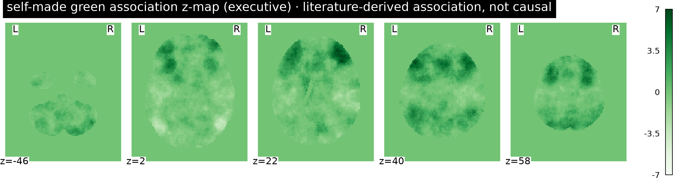
SCDWu 2025Cortical targets and EEG 10-20 fused onto one real cortex (ICBM152) — green = candidate targets, blue = EEG sites, same MNI space; the measurable side-view locator (proportions / acupoints / needles) sits below.
← back to the Scalp Fusion demo · methodology: how coordinate fusion works →
Research-use localization reference · candidate targets are literature-derived associations (not causal) · cortex + EEG use standard MNI coords; acupoints (no machine-readable 3D coords) are shown by proportional projection below · not for diagnosis · no treatment parameters.
cortical targets (size ≈ cluster)EEG 10-20 electrodesacupoints (measurable view below)
3D Brain (MNI152 · NiiVue 0.69)
Loading ICBM152 cortical mesh (~4.7 MB)…
first load 3-8s · cache hit after
6 MNI targets · loading
candidate targetconfidence halo (qualitative: approximate = tight / low = wide)secondary markeracupoint / needle point (scalp-ward)EEG 10-20
Measurable side-view locator (proportions / acupoints / needles)
Scalp 10-20 Locator
Figure: Scalp 10-20 locator · lateral view · ANTERIOR (nose) at right · blue = 10-20 reference lines · green numbered = reference points (operable) · red = caution/contraindication · brown = MS6 / cun ruler (points carry numbers — not color-dependent)
blue = 10-20 reference lines (nasion–inion · coronal, for needling localization) · green numbered dots = reference points · Acupoints: BL8 · GB10 · GB14 · GB8 · GB9 · TE23